
Wikipedia:WikiProject AP Biology Bapst 2014

- Past Related Projects: Wikipedia:WikiProject AP Biology Bapst 2012 & Wikipedia:WikiProject AP Biology 2011 & Wikipedia:WikiProject AP Biology Bapst 2013
A high school class in Maine - John Bapst Memorial High School in Bangor, Maine - will contribute images to Wikipedia article and the commons until June 6, 2014. The collective goal is to contribute excellent biology diagrams to the Commons and to corresponding Wikipedia articles. This is done as part of an Advanced Placement Biology course. The lead editor is Chris Packard. This project is inspired by the 2009 Wikipedia AP Biology Project. There are many basic and important diagrams missing from biological articles and we're doing our part to fix this.
- Students will work alone, there are 23 students so we should have 23 new images with captions and labels.
- The time frame will be three weeks.
- Students will be required to write a summary of why they select a topic; hopefully, eliminating obscure, random topic selections. They also must create labels and captions for their photos
- They may add it to encyclopedia articles.
- The best of the bunch will be submitted as Wikipedia featured pictures, see other candidates here. Featured images must be in .svg (vector) format.
Feel free to discuss this project. Please notify me of any concerns; especially if they involve the behavior of my students on Wikipedia. With a little patience, this should be an inspirational experience for all.
Goals / Motivation
- To improve the images in Wikipedia's coverage of Biology articles.
- To encourage promising students to write, create, learn, and contribute volunteer efforts through a service learning project.
- The dreaded “Research Project” is a standard hurdle for most AP Programs. Rightfully so, being that many college courses require such publications to validate your existence. This new approach to constructing a scientific document, is far more authentic and interesting. Rather than researching for a paper that is destined for the teacher's eyes and then a one way trip to the circular bin, let us contribute to the world-wide data base for others to benefit. I hope this will be an interesting and memorable project and assessment. It's funny, I can remember a number of projects and papers I wrote during my own high school experience, but I can remember no tests whatsoever.
Contributions
As you upload your projects and add them to Wikipedia please add them to the gallery below. By adding a new line which begins with the word "File" and them follows the format of my sample image. Make sure to include your caption.
-
 The above diagram is a generalized dicot seed (1) versus a generalized monocot seed (2). A. Seed Coat B. Cotyledon C. Hilum (dicots only) D. Plumule E. Radicle F. Endosperm (only in monocots)
 -
 Tadpole circulatory system. Deoxygenated blood passes near the gills and becomes reoxygenated. The blood flows through the organism until it has become depleted of oxygen once more. The blood travels in a loop that always passes through the heart once at some point. 1 - The internal gills / point where the blood is reoxygenated 2 - Point where the blood is depleted of oxygen 3 - Two chambered heart Red - Oxygenated blood. Blue - Oxygen depleted blood. The circulatory system for most fish and juvenile amphibians consists of a two chambered heart and blood vessels. The heart pumps deoxygenated blood in the blood vessels to the gills, which can be either internal or external, where it becomes stocked with oxygen. The oxygenated blood then travels through the blood vessels to all the parts of the body. When the blood gives all its oxygen to the parts, it goes back to the gills to be reoxygenated. Water, containing oxygen, flows over the gills. Since the gills in a tadpole are internal, the tadpole needs to gulp down water through its mouth. The system operates in a loop, so oxygen is always being delivered. There are thousands of blood vessels from which blood can travel. In one circuit, blood only passes through the heart once.
 -
 SAMPLE FROM 2013: 1-Water is passively transported into the roots and then into the xylem. 2-The forces of cohesion and adhesion cause the water molecules to form a column n the xylem. 3- Water leaves from the xylem into the spongy mesophyll, where it is evaporated out of the plant through the stomata.
 -
 Paramecium diagram. The parts are as follows: 1) food vacuoles 2) micronucleus 3) oral groove 4) gullet 5) anal pore 6) contractile vacuole 7) micronucleus 8) cilia.
 -
 Natural competence Caption:Key:1-Bacterial cell DNA2-Bacterial cell plasmids3-Sex pili4-Plasmid of foreign DNA from a dead cell5-Bacterial cell restriction enzyme6-Unwound foreign plasmid7-DNA ligaseI: A plasmid of foreign DNA from a dead cell is intercepted by the sex pili of a naturally competent bacterial cell.II: The foreign plasmid is transduced through the sex pili into the bacterial cell, where it is processed by bacterial cell restriction enzymes. The restriction enzymes break the foreign plasmid into a strand of nucleotides that can be added to the bacterial DNA.III: DNA ligase integrates the foreign nucleotides into the bacterial cell DNA.IV: Recombination is complete and the foreign DNA has integrated into the original bacterial cell’s DNA and will continue to be a part of it when the bacterial cell replicates next.
 -
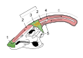 Seen here is a Parasaurolophus skull with a view into nasal cavity of the cranial crest. It is hypothesized that the Parasaurolophus pushed air through its long cranial crest to make low register sounds that could be heard for miles.
1. Nostril
2. Sinus cavity
3. Skin
4. Intra Cranial bone
5. Skull
6. Cranial crest -
 Biomagnification of toxins in the food chain of a terrestrial environment. The dots represent the organic molecules present in each trophic level. The crosses represent the mercury present in each trophic level. While dots remain relatively constant in each individual, the concentration of crosses become greater in each preceding trophic level.
 -
 This is a cross section of a normal sea urchin. The cross section is not symmetrical to show the five point radial symmetry.
1 - genital plate
2 - gonopore
3 - anus
4 - hard plate with madreporite
5 - axial gland
6 - gonad
7 - intestine
8 - ampullae
9 - test
10 - radial canal
11 - esophagus
12 - Aristotle’s lantern
13 - teeth
14 - mouth
15 - nerve ring
16 - ring canal
17 - test plates
18 - tube feet
19 - spines -
 Primary succession is the process characterized by soil and organisms becoming established in an area lacking topsoil and vegetation. The process occurs in the following steps: I. The bare rock is exposed by glaciers or volcanic eruptions. II. Lichens and moss begin to grow on the rock. III. As the lichens and moss die, they decompose, which creates the first organic soil. The rock will slowly decay, and the soil depth will increase as the organisms in the following steps die and decompose. IV. The lichens and grass grow together. V. The grasses and small herbaceous plants thrive. VI. Shrubs and bushes appear in the deeper soil, which contains more water. VII. Smaller trees develop. VIII. Larger trees and shade tolerant plants dominate the landscape. The larger plant species contribute to a faster soil growth rate because of their leaves falling to the surface floor where they decompose. Not all plant species will survive in unison. As large plants grow, they compete for sunlight and nutrients creating unfavorable environments for shade intolerant organisms.
 -
 1. Shows a plant lacking gibberellins and has a internode length of "0" as well as it is a dwarf plant. 2. Shows your average plant with a moderate amount of gibberellins and an average internode length. 3.Shows a plant with a large amount of gibberellins and so has a much longer internode length because gibberellins promotes cell division in the stem.
 -
 This is a cross section of lichen, which is an organism in a symbiotic relationship between green algae and fungus. 1. Thick layers of hyphae, called the cortex 2. Green algae 3. Loosely packed hyphae 4. Anchoring hyphae called rhizines.
 -
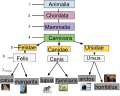 The taxonomic ranking system is used to characterize organisms. Ordered from broadest to most specific, the basic groups go as follows:1-Domain2-Kingdom3-Phylum 4-Class5-Order6-Family7-Genus8-SpeciesEach division of the ranking system separates one organism from another based on its characteristics. The organism’s scientific name is written as the genus capitalized and its species lower case. (Genus species). This is called the binomial nomenclature system developed by Carolus Linnaeus.
 -
 This Punnett square displays phenotypic incomplete dominance. Incomplete dominance is when the there is no dominance between traits, and it results in a blending of traits. In this example, the R allele for red petals blends with the white trait of the r allele. This results in the petals being pink, which is a trait of neither allele.
 -
 The glycocalyx exists in bacteria as either a capsule or a slime layer. 6 points at the glycocalyx. The difference between a capsule and a slime layer is that in a capsule polysaccharides are firmly attached to the cell wall, while in a slime layer the glycoproteins are loosely attached to the cell wall.
 -
 Male platypus reproductive system. 1.Testes 2.Epididymis 3.Bladder 4.Rectum 5.Ureter 6.Vas Deferens 7.Genito - Urinary Sinus 8.Penis enclosed in a fibrous sheath 9.Cloaca 10.Opening in the ventral wall of the cloaca for the penis.
 -
 The specific immune response of a mammal: CAPTION:
 -
 1. Ligands
2. Receptors
3. Secondary Messengers
These are examples of membrane receptors. Typically, they are proteins that are embedded in the membrane. Although there are many different ligands located outside of the cell, membrane proteins are specific, and only certain ligands will bind to each one. That is why each protein has a different ligand, and also induces a different cellular response. The response may be transcription of a gene, cell growth, or many other cellular actions. -
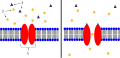 1. Ion Channel Receptor
2. Ions
3. Ligand (such as acetylcholine)
This is an example of an ion channel receptor. On the left, the channel is closed, because the ligand (dark purple triangle) has not binded to the receptor. When the ligand binds to the receptor, the channel opens, and the ions (orange circles) can freely flow through the membrane.
In a neuromuscular junction, these are used to transfer the action potential from the neuron to the muscle. The ligand is acetylcholine, and when it binds to the ion channel receptor on the membrane of the muscle, the channel opens and allows sodium ions to flow into the muscle. -
 A - Active Site
B - Allosteric Site
C - Substrate
D - Inhibitor
E - Enzyme
1. In this process, the substrate (C) binds to the enzyme (E) at the active site (A). This enzyme is functioning normally, and is not inhibited.
2. In this process, an inhibitor (D) binds to the allosteric site (B) on the enzyme (E), causing a change in the shape of the enzyme. The substrate (C) can no longer bind to the active site (A) of the enzyme. -
 1. Esophagus
2. Stomach
3.Ulcers
4.Duodenum
5.Mucosa
6.Submucosa
7.Muscle
When peptic ulcers occur, they can be found in either the lining of the stomach, the lining of the duodenum, or in both. Peptic ulcers that occur in the stomach are named gastric ulcers whereas ulcers found in the duodenum are referred to as duodenal ulcers. Peptic ulcers can be minor (they only go through the first or the second layers of the stomach), or can be considered a medical emergency (they go through every layer of the stomach or duodenum lining causing major internal bleeding). A peptic ulcer is a defect that occurs in the first part of the small intestine or in the lining of the stomach. Small ulcers may not be noticeable, whereas large ulcers are considered medical emergencies. Abdominal pain is a common symptom seen with multiple types of ulcers. Common causes of ulcers can be: bacterial infections in the stomach or duodenum, a weakening of the stomach lining caused by a continuous use of an anti inflammatory, or the common belief that peptic ulcers are caused by stress.
http://www.wisegeek.org/what-are-ulcers.htm . -
 Bergmann's Rule is an ecologic principle which states that as latitude increases the body mass of a particular species increases. The data are taken from a Swedish study investigating the size of moose as latitude increases as shows the positive relationship between the two, supporting Bergmann’s Rule. Sand, Håkan, Göran Cederlund, and Kjell Danell. "Geographical and Latitudinal Variation in Growth Patterns and Adult Body Size of Swedish Moose (Alces Alces)." Oecologia 102.4 (1995): 433-42. Web.
 -
 Male whale reproductive system: Whale penises are fully inside the body until the whale becomes erect. The testicles are inside the body at all times. 1. Genital Slit 2. Penis 3. Anus 4. Testes 5. Vas Deferens 6. Kidney 7. Diaphragm Whale penises are fully inside the body until the whale becomes erect. The testicles are inside the body at all times.
 -
 1. Tentacles 2. Oral disk 3. Contracting muscles 4. Gonads 5. Acontial filaments 6. Pedal disk 7. Ostium 8. Coelenteron. Sea anemones have soft tube like bodies. The external structure consists of the tentacles, the oral disk, and the pedal disk. The tentacles, which are covered in nematocysts, capture and transport prey to the oral disk. The oral disk serves as both the mouth and the anus. The mouth is the opening to the coelenteron, a single sac like cavity that performs all digestive functions. The pedal disk attaches the sea anemone to hard surfaces. The internal structure of a sea anemone consists of the contracting muscles, the gonads, the acontial filaments, and the ostium. The contracting muscles consist of simple longitudinal fibers that contract to move the anemone vertically.The gonads can be found in the mesentery. The ostium are where water is let in and out of the anemone. The acontial filament are found in the bottom sac section of the coelenteron. The acontial filaments are laden with nematocysts. Acontia filaments are used for protection from predators.
 -
 Euglena are unicellular, flagellate protists of the genus Euglena and kingdom Eukarya. Able to photosynthesize with their chloroplasts and also capable of accessing food from outside sources, they are both autotrophic and heterotrophic. Euglena have both plantlike and animalistic qualities: they contain chloroplasts, but have a pellicle, an envelope made up of many microtubules allowing for fluid, smooth movement in their environment. Although they are eukaryotic organisms, they reproduce asexually through binary fission. Some components of the anatomy of euglena are very unique: Euglena have two flagella, even though only one is visible outside the cell: the second flagellum does not protrude out of the reservoir. The red eyespot of a euglena filters light for the photoreceptor so that only certain wavelengths of light are able to reach the photoreceptor, allowing the euglena to “steer” itself by moving toward light in different intensities in different areas of its photoreceptor. Key: 1. Microtubules that make up the pellicle (see 9.) 2. Contractile Vacuole 3. Stigma / Eyespot 4. Flagella 5. Reservoir 6. Photoreceptor / Paraflagellar Body 7. Basal Bodies 8. Chloroplast 9.Pellicle 10. Nucleus 11. Nucleolus
 -
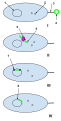
 This is a diagram showing the Green Beard Effect in action. It shows the tendency of odd, out of the norm individuals to prefer other, similarly odd individuals. This is thought to be due to the sexual reproduction mechanism and the random shuffling of genes it produces in progeny. The tendencies then must spring from the belief that mating with similar individuals with presumably similar genes will ensure your genes are passed on to the next generation to be spread further.I. 1. The Genotype shown by the two individuals here is this society’s standard - as it codes for the accepted phenotype (physical appearance); that is, light blue faces. 2. The two individuals are attracted to each other, since they share mutual characteristics and therefore most likely also share DNA sequences (genes) that code for these outward appearances. 3. The parents’ mutual genes for face color are passed on to their offspring, which facilitates the spreading of their genes to future generations, benefiting both parents. II. 1. The individual on the right possesses the common, accepted light blue face genotype (RR); however, the one on the left has a rare, generally disliked orange face genotype (rr). 2. The two organisms are repulsed by each other, since their physical characteristics are different and therefore the genes that code for these quirks.3. No children are had, seeing as both organisms would be faced with uncertainty over whether their progeny (children) would carry their genes to the next generation. III. 1. Both individuals possess the rare orange face color gene (rr) that is generally found repulsive by most members of society.2. The two organisms are attracted to one another, even though most others find the characteristic repulsive since they have the same gene for face color (rr). 3. The members pass their mutual gene for orange face color on to their offspring. This is thought to be the reason why two generally repulsive individuals would be mutually attracted to one another. The rare genotype (rr) possessed by both of the members that results in the repulsive quirks causes the odd individuals to want to reproduce together since it will be guaranteed that the rare (rr) genotype will be passed on to the next generation and spread.
 -
 The competitive exclusion principle states that no two species that fill a similar role in an ecosystem can occupy the same area. For example, Chthamalus and Balanus (two species of barnacle) cannot live in the same tidal zone. If only Chthamalus is present, it can grow all the way down to the low tide area (where Balanus normally is). If only Balanus is there, it can grow into the high tide area (Chthamalus’s normal territory). If both Chthamalus and Balanus are living together, Chthamalus is in the high tide and Balanus is in the low tide. Because both species occupy similar niches, they cannot live in the same area because they compete for the same resources. I. Chthamalus and Balanus together II. Balanus by itself III. Chthamalus by itself Labels: 1. Chthamalus 2. Balanus 3. Area of Competition 4. High Tide 5. Low Tide Source: Mader, Sylvia S. Biology. 10th ed. Boston: WCB/McGraw-Hill, 1998. 843. Print.


Contributors
Add your user name here following my example. Just add this template with your username instead of the line: {{user|username}} and then, if your username is not identifiable, your real first name.
- Ralph Walterberg (talk · contribs) - Braydon - Myelin Sheath Cross Section
- Earthdirt (talk · contribs) - Chris (AKA Mr. Packard) - IMAGE TOPIC NAME HERE
- Jofalee (talk · contribs) - Lee - Gibberlins
- cbenson1919 (talk · contribs) - Courtney- Monocot Seed vs. Dicot Seed
- tessaelilley (talk · contribs) - Tessa- Taxonomic rank
- nmccarthy16 (talk · contribs) - Noah- Bergmann's Rule
- Spencerbaron (talk · contribs) - Spencer- "Incomplete dominance"
- opellegrini15 (talk · contribs) - Olivia - "Juvenile Amphibian Circulatory System"
- jbell16 (talk · contribs) Jocelyn- Competitive Exclusion Principle
- erinlandry (talk · contribs) - Erin - "Sea Urchin Anatomy"
- JanisIan15 (talk · contribs) - Cooper - ""Specific Immune Response""
- jsun15 (talk · contribs) - Bob - ""Glycocalyx ""
- klarochelle16 (talk · contribs) - Katherine - Natural Competence
- sballesteros15 (talk · contribs) - Samantha - Biomagnification
- ImaSmurf (talk · contribs) - Isaiah - Green Beard Effect
- Whizzy1999 (talk · contribs) - Isaac - Receptor (biochemistry)
- joshfn (talk · contribs)- Joshua Nosel - Primary Succe ssion
- jdurant (talk · contribs) - Jasmine - Lichen
- ciarachristina (talk · contribs) - Ciara Petrello - Paramecium Diagram
- lydiakurkoski (talk · contribs) Lydia Kurkoski- Sea Anemone Diagram
- rachaelgraves (talk · contribs) -Rachael Graves- Peptic ulcer
- chillery16 (talk · contribs) - Caitlin - "Euglena"
- jgagnon14 (talk · contribs) - Justin- Whale sex Organs
- esouc32 (talk · contribs) - Elliott - Male Platypus Reproductive System
- Jdistefano14 (talk · contribs) - Joe - Parasaurolophus Cranial Crest
Uploading
In order to complete the assignment and reap all the benefits of your hard work (such as a good grade) you MUST complete all of the following steps. If you need help, just ask.
How to, step by step
Step 1: Create a Wikipedia Global account by clicking "Login/create account" in the upper right hand corner of this page.
Step 2: Click here to use the WikiCommons File Upload Wizard
Step 3: If you didn't do it in the Wizard, categorize your image by adding a one or more [[Category:_______]] tags at the bottom of the page (fill in the name of the category in the _______.) You might use Category:Biology diagrams or something more specific like Category:Molecular biology or something else appropriate.
Step 4: If you didn't do it in the Wizard you should also now add your labels and your caption information in the description to your upload page in the Commons.
Step 5: Your image is now available in all Wiki Projects, including Wikipedia. So let's add it to the article! Go to the article you want to add your donated image to. In the top of the section of the article or the subheading you want to add the image to add something like this:
[[File:MY IMAGE NAME.png|right|thumb|200px|The [[caption]] of '''my image'''.]]
That's not too hard is it? For your caption you'll need to follow Wikipedia style and use some mark up to do this - it's kind of like a micro-essay. The [[ ]] creates a link to the given page on Wikipedia and the ''' ''' make the word bold, in Wikipedia it's appropriate to bold the title of the article the first time it's used in the text or in a caption."
Step 6: Wow you've done it! Now you just have to turn in your work by adding it to gallery in the section above here called "Contributions". Just follow the model I provided in the first entry. Your caption will likely have to be shorter than your description, see the style advice below.
Style guides
To get past the stumbling blocks of editing Wikipedia, articles will have to conform to the Wikipedia style guides. The largest barriers are:
- Wikipedia:Manual of Style/Images - The basic overview of images (the Wikipedia:Picture tutorial is also useful.
- Wikipedia:Manual of Style/Captions - Writing a good caption may be harder than you think.
- Wikipedia:Copyrights - Make sure to post a license on your image which releases all copyrights and makes it free use image AND don't use images from anywhere except the Commons if your image integrates other images.
- Wikipedia:File names - Pick the right name for your file.
- Wikipedia:Preparing images for upload - Pick the right file type (images created using entirely Google Draw should be saved as .SVG, whereas most other images you make will be saved as a .PNG in rare cases an a .JPG or .JPEG can be used)
- Wikipedia:Uploading images or WikiCommons Uploading Images - Do it right the first time (or just use the Wizard).
- Wikipedia:Ten things you may not know about images on Wikipedia - Kind of interesting.
You can always ask for help at:
Writing a good image caption
There are several criteria for a good caption. A good caption:
- clearly identifies the subject of the picture, without detailing the obvious.
- is succinct (that means short).
- establishes the picture's relevance to the article.
- provides context for the picture.
- draws the reader into the article.
Different people read articles different ways. Some people start at the top and read each word until the end. Others read the first paragraph and scan through for other interesting information, looking especially at pictures and captions. For those readers, even if the information is adjacent in the text, they will not find it unless it is in the caption—but do not tell the whole story in the caption—use the caption to make the reader curious about the subject.
Another way of approaching the job: imagine you're giving a lecture based on the encyclopedia article, and you are using the image to illustrate the lecture. What would you say while attention is on the image? What do you want your audience to notice in the image, and why? Corollary: if you have got nothing to say, then the image probably does not belong in the article.
Images for the lead
It is very common to use an appropriate representative image for the lead of an article, often as part of an infobox. The image helps to provide a visual association for the topic, and allows readers to quickly assess if they have arrived at the right page. For most topics, the selection of a lead image is plainly obvious: a photograph or artistic work of a person, photographs of a city, or a cover of a book or album, to name a few.
Image selection for other topics may be more difficult and several possible choices could be made. While Wikipedia is not censored, as outlined in the above section on offensive images, the selection of the lead image should be made with some care with respect to this advice. Lead images are loaded and shown upon navigating to the page, and are one of the first things that readers will see. Editors should avoid using images that readers would not have expected to see when navigating to the page. Unlike other content on a page that falls below the lead, the lead image should be chosen with these considerations in mind.
Some advice on selecting a lead image include the following:
- Lead images should be images that are natural and appropriate visual representations of the topic; they not only should be illustrating the topic specifically, but should also be the type of image that is used for similar purposes in high-quality reference works, and therefore what our readers will expect to see. Lead images are not required, and not having a lead image may be the best solution if there is no easy representation of the topic.
- Lead images should be selected to be of least shock value; if an alternative image exists that still is an accurate representation of the topic but without shock value, it should always be preferred. For example, using an image of deportees being subjected to selection as the lead image at this version of Holocaust is far preferable to the appropriate images that appear later in the article that show the treatment of the prisoners or corpses from the camps.
- Sometimes it is impossible to avoid the use of a lead image with perceived shock value if the topic itself is of that nature, for example in articles on various parts of human genitalia. It should be anticipated, through Wikipedia:Content disclaimer, that readers will be aware they will be exposed to potentially shocking images when navigating to articles on such topics.
Planning and resources
- Wikipedia tutorials for beginners
- Editing commands cheatsheet
- Getting started
- The perfect article
- Assessment
- Article development
- Peer Review
- [[Active gif creator]]
Talk pages
These are places where you can leave and receive messages and questions, every page has one. Whenever you edit these pages, make sure that you are signed in. Also, add four tildes ~~~~ to the end of all comments you make on talk pages. This will let people know who is talking.