1agr: COMPLEX OF ALF4-ACTIVATED GI-ALPHA-1 WITH RGS4
1as0: GTP-GAMMA-S BOUND G42V GIA1
1as2: GDP+PI BOUND G42V GIA1
1as3: GDP BOUND G42V GIA1
1bh2: A326S MUTANT OF AN INHIBITORY ALPHA SUBUNIT
1bof: GI-ALPHA-1 BOUND TO GDP AND MAGNESIUM
1cip: GI-ALPHA-1 SUBUNIT OF GUANINE NUCLEOTIDE-BINDING PROTEIN COMPLEXED WITH A GTP ANALOGUE
1gdd: TERTIARY AND QUATERNARY STRUCTURAL CHANGES IN GIA1 INDUCED BY GTP HYDROLYSIS
1gfi: STRUCTURES OF ACTIVE CONFORMATIONS OF GI ALPHA 1 AND THE MECHANISM OF GTP HYDROLYSIS
1gg2: G PROTEIN HETEROTRIMER MUTANT GI_ALPHA_1(G203A) BETA_1 GAMMA_2 WITH GDP BOUND
1gia: STRUCTURE OF ACTIVE CONFORMATIONS OF GIA1 AND THE MECHANISM OF GTP HYDROLYSIS
1gil: STRUCTURE OF ACTIVE CONFORMATIONS OF GIA1 AND THE MECHANISM OF GTP HYDROLYSIS
1git: STRUCTURE OF GTP-BINDING PROTEIN
1gp2: G PROTEIN HETEROTRIMER GI_ALPHA_1 BETA_1 GAMMA_2 WITH GDP BOUND
1kjy: Crystal Structure of Human G[alpha]i1 Bound to the GoLoco Motif of RGS14
1svk: Structure of the K180P mutant of Gi alpha subunit bound to AlF4 and GDP
1svs: Structure of the K180P mutant of Gi alpha subunit bound to GppNHp.
1y3a: Structure of G-Alpha-I1 bound to a GDP-selective peptide provides insight into guanine nucleotide exchange
2g83: Structure of activated G-alpha-i1 bound to a nucleotide-state-selective peptide: Minimal determinants for recognizing the active form of a G protein alpha subunit
2gtp: Crystal structure of the heterodimeric complex of human RGS1 and activated Gi alpha 1
2hlb: A Structural Basis for Nucleotide Exchange on G-alpha-i Subunits and Receptor Coupling Specificity
2ihb: Crystal structure of the heterodimeric complex of human RGS10 and activated Gi alpha 3
2ik8: Crystal structure of the heterodimeric complex of human RGS16 and activated Gi alpha 1
2ode: Crystal structure of the heterodimeric complex of human RGS8 and activated Gi alpha 3

 1agr: COMPLEX OF ALF4-ACTIVATED GI-ALPHA-1 WITH RGS4
 1as0: GTP-GAMMA-S BOUND G42V GIA1
 1as2: GDP+PI BOUND G42V GIA1
 1as3: GDP BOUND G42V GIA1
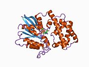 1bh2: A326S MUTANT OF AN INHIBITORY ALPHA SUBUNIT
 1bof: GI-ALPHA-1 BOUND TO GDP AND MAGNESIUM
 1cip: GI-ALPHA-1 SUBUNIT OF GUANINE NUCLEOTIDE-BINDING PROTEIN COMPLEXED WITH A GTP ANALOGUE
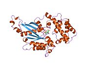 1gdd: TERTIARY AND QUATERNARY STRUCTURAL CHANGES IN GIA1 INDUCED BY GTP HYDROLYSIS
 1gfi: STRUCTURES OF ACTIVE CONFORMATIONS OF GI ALPHA 1 AND THE MECHANISM OF GTP HYDROLYSIS
 1gg2: G PROTEIN HETEROTRIMER MUTANT GI_ALPHA_1(G203A) BETA_1 GAMMA_2 WITH GDP BOUND
 1gia: STRUCTURE OF ACTIVE CONFORMATIONS OF GIA1 AND THE MECHANISM OF GTP HYDROLYSIS
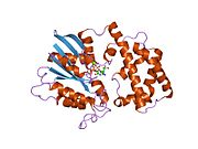 1gil: STRUCTURE OF ACTIVE CONFORMATIONS OF GIA1 AND THE MECHANISM OF GTP HYDROLYSIS
 1git: STRUCTURE OF GTP-BINDING PROTEIN
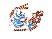 1gp2: G PROTEIN HETEROTRIMER GI_ALPHA_1 BETA_1 GAMMA_2 WITH GDP BOUND
![1kjy: Crystal Structure of Human G[alpha]i1 Bound to the GoLoco Motif of RGS14](//upload.wikimedia.org/wikipedia/commons/thumb/e/ea/PDB_1kjy_EBI.jpg/180px-PDB_1kjy_EBI.jpg) 1kjy: Crystal Structure of Human G[alpha]i1 Bound to the GoLoco Motif of RGS14
1kjy: Crystal Structure of Human G[alpha]i1 Bound to the GoLoco Motif of RGS14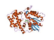 1svk: Structure of the K180P mutant of Gi alpha subunit bound to AlF4 and GDP
 1svs: Structure of the K180P mutant of Gi alpha subunit bound to GppNHp.
 1y3a: Structure of G-Alpha-I1 bound to a GDP-selective peptide provides insight into guanine nucleotide exchange
 2g83: Structure of activated G-alpha-i1 bound to a nucleotide-state-selective peptide: Minimal determinants for recognizing the active form of a G protein alpha subunit
 2gtp: Crystal structure of the heterodimeric complex of human RGS1 and activated Gi alpha 1
 2hlb: A Structural Basis for Nucleotide Exchange on G-alpha-i Subunits and Receptor Coupling Specificity
 2ihb: Crystal structure of the heterodimeric complex of human RGS10 and activated Gi alpha 3
 2ik8: Crystal structure of the heterodimeric complex of human RGS16 and activated Gi alpha 1
 2ode: Crystal structure of the heterodimeric complex of human RGS8 and activated Gi alpha 3


![1kjy: Crystal Structure of Human G[alpha]i1 Bound to the GoLoco Motif of RGS14](/File:PDB_1kjy_EBI.jpg)


