Antibiotic sensitivity testing: Difference between revisions
typo |
→Clinical practice: not needed. The Talk Page should be used rather than lengthy edit summaries |
||
| Line 29: | Line 29: | ||
Often clinical specimens are sent to the [[medical laboratory|clinical laboratory]] for culture and sensitivity, which is culture and antibiotic sensitivity testing offered as one combined service.<ref name="Guiliano2019" /> This is done through collecting samples from affected body sites, preferably before antibiotics are given. For example, a person in an [[intensive care]] unit may develop a [[hospital-acquired pneumonia]]. There is a chance the causal bacteria may be different to [[community-acquired pneumonia]].<ref name="pmid30596308">{{cite journal |vauthors=Peyrani P, Mandell L, Torres A, Tillotson GS |title=The burden of community-acquired bacterial pneumonia in the era of antibiotic resistance |journal=Expert Review of Respiratory Medicine |volume=13 |issue=2 |pages=139–152 |date=February 2019 |pmid=30596308 |doi=10.1080/17476348.2019.1562339 |url=}}</ref> Treatment is generally started empirically, on the basis of surveillance data about the local common bacterial causes. This first treatment, based on statistical information about former patients, and aimed at a large group of potentially involved microbes, is called [[empirical treatment]].{{sfn|Burnett|2005|pp=167–171}} |
Often clinical specimens are sent to the [[medical laboratory|clinical laboratory]] for culture and sensitivity, which is culture and antibiotic sensitivity testing offered as one combined service.<ref name="Guiliano2019" /> This is done through collecting samples from affected body sites, preferably before antibiotics are given. For example, a person in an [[intensive care]] unit may develop a [[hospital-acquired pneumonia]]. There is a chance the causal bacteria may be different to [[community-acquired pneumonia]].<ref name="pmid30596308">{{cite journal |vauthors=Peyrani P, Mandell L, Torres A, Tillotson GS |title=The burden of community-acquired bacterial pneumonia in the era of antibiotic resistance |journal=Expert Review of Respiratory Medicine |volume=13 |issue=2 |pages=139–152 |date=February 2019 |pmid=30596308 |doi=10.1080/17476348.2019.1562339 |url=}}</ref> Treatment is generally started empirically, on the basis of surveillance data about the local common bacterial causes. This first treatment, based on statistical information about former patients, and aimed at a large group of potentially involved microbes, is called [[empirical treatment]].{{sfn|Burnett|2005|pp=167–171}} |
||
Before starting this treatment, the physician will collect a sample from the site of a suspected infection: a [[blood culture]] sample when bacteria possibly have invaded the bloodstream, a [[sputum]] sample in the case of a ventilator associated pneumonia, and a [[urine]] sample in the case of a urinary tract infection. These samples are transferred to the [[microbiology]] laboratory where they are added to [[microbiological culture|culture media]].{{sfn|Burnett|2005|pp=135–144}} Microscopy is not useful for all samples because often the normal bacterial flora cannot be distinguished from pathogens.{{sfn | Pommerville | 2018 | p=632}} Additionally, when reported, a decision must be made for some bacteria such as [[staphylococcus epidermidis]], as to whether they are the cause of an infection, or simply [[Commensalism|commensual]] bacteria or contaminants.<ref name="Guiliano2019" /><ref>{{cite journal |last1=Becker |first1=Karsten |last2=Heilmann |first2=Christine |last3=Peters |first3=Georg |title=Coagulase-Negative Staphylococci |journal=Clinical Microbiology Reviews |date=2 October 2014 |volume=27 |issue=4 |pages=870–926 |doi=10.1128/CMR.00109-13 |
Before starting this treatment, the physician will collect a sample from the site of a suspected infection: a [[blood culture]] sample when bacteria possibly have invaded the bloodstream, a [[sputum]] sample in the case of a ventilator associated pneumonia, and a [[urine]] sample in the case of a urinary tract infection. These samples are transferred to the [[microbiology]] laboratory where they are added to [[microbiological culture|culture media]].{{sfn|Burnett|2005|pp=135–144}} Microscopy is not useful for all samples because often the normal bacterial flora cannot be distinguished from pathogens.{{sfn | Pommerville | 2018 | p=632}} Additionally, when reported, a decision must be made for some bacteria such as [[staphylococcus epidermidis]], as to whether they are the cause of an infection, or simply [[Commensalism|commensual]] bacteria or contaminants.<ref name="Guiliano2019" /><ref>{{cite journal |last1=Becker |first1=Karsten |last2=Heilmann |first2=Christine |last3=Peters |first3=Georg |title=Coagulase-Negative Staphylococci |journal=Clinical Microbiology Reviews |date=2 October 2014 |volume=27 |issue=4 |pages=870–926 |doi=10.1128/CMR.00109-13|url=https://cmr.asm.org/content/27/4/870}}</ref> |
||
When antibiotic sensitivity testing is reported, it will provide useful information about the organisms present in the sample, and which antibiotics will be effective.<ref name="Guiliano2019" /> Antibiotic sensitivity testing is done in a laboratory ([[in vitro]]), but the correlation of this testing to the sensitivity of the antibiotics in a person ([[in vivo]]) is often high enough for the test to be clinically useful.{{sfn|Burnett|2005|p=168}} |
When antibiotic sensitivity testing is reported, it will provide useful information about the organisms present in the sample, and which antibiotics will be effective.<ref name="Guiliano2019" /> Antibiotic sensitivity testing is done in a laboratory ([[in vitro]]), but the correlation of this testing to the sensitivity of the antibiotics in a person ([[in vivo]]) is often high enough for the test to be clinically useful.{{sfn|Burnett|2005|p=168}} |
||
Revision as of 17:28, 6 July 2020

Antibiotic sensitivity testing or antibiotic susceptibility testing is the measurement of the susceptibility of bacteria to antibiotics. It is used because bacteria may have resistance to some antibiotics. Knowledge about what antibiotics a bacterium is sensitive to can change the choice of antibiotics from empiric therapy to directed therapy.[1]
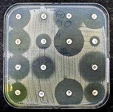
Sensitivity testing usually occurs in a laboratory setting, and may be based on culture methods that exposure bacteria to antibiotics, or genetic methods that test to see if a bacterium possesses genes that confer resistance. Culture methods often involve measuring the diameter of the zones of inhibition on agar culture dishes of bacterial growth around paper discs that are impregnated with antibiotics. The minimum inhibitory concentration of the antibiotic can be determined from this.
Uses
In clinical medicine, antibiotics are most frequently prescribed on the basis of a person's symptoms and general guidelines, called empiric therapy.[1] These are based around knowledge about what bacteria cause an infection, and also what antibiotics bacteria may be sensitive or resistant to in a geographical area.[1] For example, a simple urinary tract infections might be treated with trimethoprim/sulfamethoxazole.[2] This is because Escherichia coli is the most likely causative bacterium, and may be sensitive to that combination antibiotic.[2] However, bacteria can be resistant to several classes of antibiotics.[2] This resistance might be because of the type of bacteria,[2] because of resistance following past exposure to with antibiotics,[2] or because resistance may be transmitted from other sources such as plasmids.[3] Antibiotic sensitivity testing provides information about what antibiotics are more likely to be successful and are used in this context to provide information about what antibiotics should be used.[4][1]
Antibiotic sensitivity testing is also conducted at a population level in some countries as a form of screening.[5] This is to assess the background rates of resistance to antibiotics (for example with Methicillin-resistant Staphylococcus aureus), and may influence guidelines and public health measures.[5]
Methods
Testing for antibiotic sensitivity usually occurs in a laboratory setting.[6] Methods of testing include:
- Those based on exposing bacteria to antibiotics:
- Kirby-Bauer method. Small paper discs containing antibiotics are placed onto a plate upon which bacteria are growing. If the bacteria are sensitive to the antibiotic, a clear ring, or zone of inhibition, is seen around the wafer indicating poor growth.[7] Müeller-Hinton agar is frequently used in this antibiotic susceptibility test.[8]
- Automated methods [9]
- An Etest (also based on antibiotic diffusion) [9]
- Agar and Broth dilution methods for minimum inhibitory concentration (MIC) determination.[10][11]
- Genetic testing, such as via polymerase chain reaction, DNA microarray, DNA chips, and loop-mediated isothermal amplification, which may be used to detect whether bacteria possess genes which confer antibiotic resistance.[4][12]
Reporting
The results of the testing are reported as a table, sometimes called an antibiogram.[13] Bacteria might be marked as sensitive, resistant, or having intermediate resistance to an antibiotic.[6] Specific patterns pf drug resistance or multi drug resistance may be noted, such as the presence of an extended-spectrum beta lactamase.[6]
The sensitive, resistant or intermediate resistance to antibiotics is reported based on the minimum inhibitory concentration. It is compared to known values for a given bacterium and antibiotic.[6] For example, Streptococcus pneumoniae isolates are considered susceptible to penicillin if MICs are ≤0.06 μg/ml, intermediate if MICs are 0.12 to 1 μg/ml, and resistant if MICs are ≥2 μg/ml.[14][15] Such information may be useful to the clinician, who can change the empirical treatment, to a more custom-tailored treatment that is directed only at the causative bacterium.[1] Sometimes, whether an antibiotic is marked as resistant is also based on bacterial characteristics that are associated with known methods of resistance such as the potential for beta lactamase production.[16][6]
Clinical practice

Ideal antibiotic therapy is based on determining the causal agent and its antibiotic sensitivity. Empirical treatment is often started before laboratory microbiological reports are available when treatment should not be delayed due to the seriousness of the disease. The effectiveness of individual antibiotics varies with the anatomical site of the infection, the ability of the antibiotic to reach the site of infection, and the ability of the bacteria to resist or inactivate the antibiotic.[18]
Often clinical specimens are sent to the clinical laboratory for culture and sensitivity, which is culture and antibiotic sensitivity testing offered as one combined service.[6] This is done through collecting samples from affected body sites, preferably before antibiotics are given. For example, a person in an intensive care unit may develop a hospital-acquired pneumonia. There is a chance the causal bacteria may be different to community-acquired pneumonia.[19] Treatment is generally started empirically, on the basis of surveillance data about the local common bacterial causes. This first treatment, based on statistical information about former patients, and aimed at a large group of potentially involved microbes, is called empirical treatment.[20]
Before starting this treatment, the physician will collect a sample from the site of a suspected infection: a blood culture sample when bacteria possibly have invaded the bloodstream, a sputum sample in the case of a ventilator associated pneumonia, and a urine sample in the case of a urinary tract infection. These samples are transferred to the microbiology laboratory where they are added to culture media.[21] Microscopy is not useful for all samples because often the normal bacterial flora cannot be distinguished from pathogens.[22] Additionally, when reported, a decision must be made for some bacteria such as staphylococcus epidermidis, as to whether they are the cause of an infection, or simply commensual bacteria or contaminants.[6][23]
When antibiotic sensitivity testing is reported, it will provide useful information about the organisms present in the sample, and which antibiotics will be effective.[6] Antibiotic sensitivity testing is done in a laboratory (in vitro), but the correlation of this testing to the sensitivity of the antibiotics in a person (in vivo) is often high enough for the test to be clinically useful.[24]
Further research
Point-of-care testing is being developed to speed up the time for testing, and to help practitioners avoid prescribing unnecessary antibiotics in the style of precision medicine.[25] Traditional techniques typically take 12 to 48 hours,[26] to up to five days.[6] In contrast, rapid testing using molecular diagnostics is defined as "being feasible within an 8-h(our) working shift".[26] Progress has been slow due to a range of reasons including cost and regulation.[27]
As of 2017, point-of-care resistance diagnostics was available for methicillin-resistant Staphylococcus aureus (MRSA), rifampin-resistant Mycobacterium tuberculosis (TB), and Vancomycin-resistant enterococci (VRE) through GeneXpert by molecular diagnostics company Cepheid.[28]
See also
Bibliography
- Burnett, David (2005). The science of laboratory diagnosis. Chichester, West Sussex, England Hoboken, NJ: Wiley. ISBN 978-0-470-85912-4. OCLC 56650888.
- Pommerville, Jeffrey (2018). Fundamentals of microbiology. Burlington, MA: Jones & Bartlett Learning. ISBN 978-1-284-10095-2. OCLC 979994356.
References
- ^ a b c d e Leekha, Surbhi; Terrell, Christine L.; Edson, Randall S. (February 2011). "General Principles of Antimicrobial Therapy". Mayo Clinic Proceedings. 86 (2): 156–167. doi:10.4065/mcp.2010.0639.
Once microbiology results have helped to identify the etiologic pathogen and/or antimicrobial susceptibility data are available, every attempt should be made to narrow the antibiotic spectrum. This is a critically important component of antibiotic therapy because it can reduce cost and toxicity and prevent the emergence of antimicrobial resistance in the community
- ^ a b c d e Kang CI, Kim J, Park DW, Kim BN, Ha US, Lee SJ, Yeo JK, Min SK, Lee H, Wie SH (March 2018). "Clinical Practice Guidelines for the Antibiotic Treatment of Community-Acquired Urinary Tract Infections". Infection & Chemotherapy. 50 (1): 67–100. doi:10.3947/ic.2018.50.1.67. PMC 5895837. PMID 29637759.
- ^ Partridge SR, Kwong SM, Firth N, Jensen SO (October 2018). "Mobile Genetic Elements Associated with Antimicrobial Resistance". Clinical Microbiology Reviews. 31 (4). doi:10.1128/CMR.00088-17. PMC 6148190. PMID 30068738.
- ^ a b Khan, Zeeshan A.; Siddiqui, Mohd F.; Park, Seungkyung (3 May 2019). "Current and Emerging Methods of Antibiotic Susceptibility Testing". Diagnostics. 9 (2): 49. doi:10.3390/diagnostics9020049.
{{cite journal}}: CS1 maint: unflagged free DOI (link) - ^ a b Molton, James S.; Tambyah, Paul A.; Ang, Brenda S. P.; Ling, Moi Lin; Fisher, Dale A. (2013-05-01). Weinstein, Robert A. (ed.). "The Global Spread of Healthcare-Associated Multidrug-Resistant Bacteria: A Perspective From Asia". Clinical Infectious Diseases. 56 (9): 1310–1318. doi:10.1093/cid/cit020. ISSN 1058-4838.
- ^ a b c d e f g h i Giuliano, C; Patel, CR; Kale-Pradhan, PB (April 2019). "A Guide to Bacterial Culture Identification And Results Interpretation". P & T : a peer-reviewed journal for formulary management. 44 (4): 192–200. PMID 30930604.
- ^ "Bauer-Kirby disk Diffusion". www.uphs.upenn.edu.
- ^ "Testing the Effectiveness of Antimicrobials | Microbiology". courses.lumenlearning.com. Retrieved 2019-02-28.
- ^ a b Burnett 2005, p. 169.
- ^ "Antibiotic Sensitivity Testing". October 28, 2008.
- ^ Reller, L. Barth; Weinstein, Melvin; Jorgensen, James H.; Ferraro, Mary Jane (December 1, 2009). "Antimicrobial Susceptibility Testing: A Review of General Principles and Contemporary Practices". Clinical Infectious Diseases. 49 (11): 1749–1755. doi:10.1086/647952 – via academic.oup.com.
- ^ Poirel L, Jayol A, Nordmann P (April 2017). "Polymyxins: Antibacterial Activity, Susceptibility Testing, and Resistance Mechanisms Encoded by Plasmids or Chromosomes". Clinical Microbiology Reviews. 30 (2): 557–596. doi:10.1128/CMR.00064-16. PMC 5355641. PMID 28275006.
- ^ "Medical Definition of ANTIBIOGRAM". www.merriam-webster.com. Retrieved 2020-07-05.
- ^ Jacobs MR, Bajaksouzian S, Palavecino-Fasola EL, Holoszyc HM, Appelbaum PC (January 1998). "Determination of penicillin MICs for Streptococcus pneumoniae by using a two- or three-disk diffusion procedure". Journal of Clinical Microbiology. 36 (1): 179–83. PMC 124830. PMID 9431943.
- ^ Goldsmith CE, Moore JE, Murphy PG (December 1997). "Pneumococcal resistance in the UK". The Journal of Antimicrobial Chemotherapy. 40 Suppl A: 11–8. doi:10.1093/jac/40.suppl_1.11. PMID 9484868.
- ^ Winstanley T, Courvalin P (July 2011). "Expert systems in clinical microbiology". Clinical Microbiology Reviews. 24 (3): 515–56. doi:10.1128/CMR.00061-10. PMC 3131062. PMID 21734247.
- ^ Kirby-Bauer Disk Diffusion Susceptibility Test Protocol Archived 26 June 2011 at the Wayback Machine, Jan Hudzicki, ASM
- ^ Burnett 2005, p. 167.
- ^ Peyrani P, Mandell L, Torres A, Tillotson GS (February 2019). "The burden of community-acquired bacterial pneumonia in the era of antibiotic resistance". Expert Review of Respiratory Medicine. 13 (2): 139–152. doi:10.1080/17476348.2019.1562339. PMID 30596308.
- ^ Burnett 2005, pp. 167–171.
- ^ Burnett 2005, pp. 135–144.
- ^ Pommerville 2018, p. 632.
- ^ Becker, Karsten; Heilmann, Christine; Peters, Georg (2 October 2014). "Coagulase-Negative Staphylococci". Clinical Microbiology Reviews. 27 (4): 870–926. doi:10.1128/CMR.00109-13.
- ^ Burnett 2005, p. 168.
- ^ "Diagnostics Are Helping Counter Antimicrobial Resistance, But More Work Is Needed". MDDI Online. 2018-11-20. Retrieved 2018-12-02.
- ^ a b van Belkum A, Bachmann TT, Lüdke G, Lisby JG, Kahlmeter G, Mohess A, Becker K, Hays JP, Woodford N, Mitsakakis K, Moran-Gilad J, Vila J, Peter H, Rex JH, Dunne WM (January 2019). "Developmental roadmap for antimicrobial susceptibility testing systems". Nature Reviews. Microbiology. 17 (1): 51–62. doi:10.1038/s41579-018-0098-9. PMID 30333569.
- ^ "Progress on antibiotic resistance". Nature. 562 (7727): 307. October 2018. doi:10.1038/d41586-018-07031-7. PMID 30333595.
- ^ McAdams, David (January 2017). "Resistance diagnosis and the changing epidemiology of antibiotic resistance". Annals of the New York Academy of Sciences. 1388 (1): 5–17. doi:10.1111/nyas.13300. ISSN 0077-8923. PMID 28134444.
External links
- Research Data
- Raw Data
- Antibiogram technique video (diffusion method)
