
Search results
There is a page named "Polar body" on Wikipedia
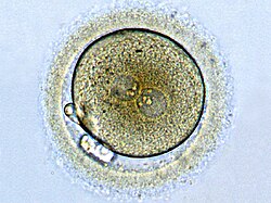
 A polar body is a small haploid cell that is formed at the same time as an egg cell during oogenesis, but generally does not have the ability to be fertilized...9 KB (1,181 words) - 06:43, 5 July 2024
 Twin (redirect from Polar body twin)that it was probably a polar body twin. The authors were unable to predict whether a healthy fetus could result from a polar body twinning. However, a study...86 KB (9,435 words) - 06:30, 28 March 2025
 Primary Oocyte —(Meiosis I)—> First Polar body (Discarded afterward) + Secondary oocyte —(Meiosis II)—> Second Polar Body (Discarded afterward) + Ovum Oocyte...30 KB (3,522 words) - 15:45, 21 March 2025
 Nondisjunction (section Polar body diagnosis)meiosis II. In oocytes, one sister chromatid is segregated into the second polar body, while the other stays inside the egg. During spermatogenesis, each meiotic...25 KB (2,857 words) - 18:11, 18 May 2024
- Polar body biopsy is the sampling of a polar body of an oocyte. It was first applied clinically in humans in 1987 after extensive animal studies. A polar...7 KB (880 words) - 22:16, 24 December 2023
 composition for a body of ice to be termed a polar ice cap, nor any geological requirement for it to be over land, but only that it must be a body of solid phase...13 KB (1,512 words) - 22:23, 11 February 2025
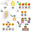 report in 2001 on worldwide use of PGD since 1990 reported that embryo or polar body biopsy had occurred in more than 3000 clinical cycles, with a 24% pregnancy...112 KB (14,647 words) - 11:24, 19 February 2025
- A polar orbit is one in which a satellite passes above or nearly above both poles of the body being orbited (usually a planet such as the Earth, but possibly...3 KB (416 words) - 05:52, 10 February 2025
 {\displaystyle K} is a bounded convex body containing the origin O {\displaystyle O} in its interior, the polar body K ∗ {\displaystyle K^{*}} is { u : ⟨...4 KB (481 words) - 04:14, 19 October 2024
 chromosome) and extrude its first polar body. The oocyte will only continue through meiosis and extrude its second polar body once it is fertilized. Ovulation...9 KB (1,039 words) - 05:47, 27 February 2025
 with bodies moving around a central point, or phenomena originating from a central point, are often simpler and more intuitive to model using polar coordinates...49 KB (6,702 words) - 11:36, 26 March 2025
 the four meiotic products are typically eliminated by extrusion into polar bodies, and only one cell develops to produce an ovum. Because the number of...76 KB (8,978 words) - 15:08, 30 March 2025
 polar body and a large secondary oocyte. The secondary oocyte undergoes meiotic division II and that results in the formation of a second small polar...31 KB (4,040 words) - 18:38, 4 February 2025
 becomes one haploid ovum and typically two polar bodies, however one may later divide to form a third polar body. In a male, meiosis starts with one diploid...10 KB (1,340 words) - 23:42, 12 August 2023
- Polar cell may refer to: Polar cells, a constituent of atmospheric circulation Polar body, a smaller cell by-product of egg formation in some animal species...196 bytes (58 words) - 19:17, 29 December 2019
- in all (4C). When meiosis I is completed, one secondary oocyte and one polar body is created. Primary oocytes have been created in late fetal life. This...4 KB (382 words) - 20:46, 30 May 2024
 and the male pronucleus. The other product of meiosis is the second polar body with only chromosomes but no ability to replicate or survive. In the fertilized...9 KB (879 words) - 12:58, 31 March 2025
 The polar bear (Ursus maritimus) is a large bear native to the Arctic and nearby areas. It is closely related to the brown bear, and the two species can...97 KB (12,613 words) - 00:15, 19 March 2025
- facultative parthenote reptiles is terminal fusion, in which a haploid polar body produced as a byproduct of normal female meiosis fuses with the egg cell...23 KB (2,749 words) - 13:38, 24 December 2024
- meiosis can complete resulting in the secondary oocyte and the first polar body. The secondary oocyte can later be fertilized with the male sperm. Sexual...9 KB (984 words) - 15:11, 1 March 2025


- English Wikipedia has an article on: polar body Wikipedia polar body (plural polar bodies) (biology) One of the small cells that are by-products of the
- Volume 8 April 1876 (1876) The Polar Glaciers I by C. C. Merriman 591327Popular Science Monthly Volume 8 April 1876 — The Polar Glaciers I1876C. C. Merriman
- [Asking him if he'll come out to his parents.] What about you, Bi-Polar Bear? Bi-Polar Bear: I already told my father, he was totally shocked... and so
- organisms. It consists of an oxygen atom connected to two hydrogen atoms by polar covalent bonds. It is considered to be the universal solvent for many reasons