
Search results
There is a page named "File:Inverted microscope.jpg" on Wikipedia
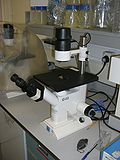 (2295368 bytes) By Richard Wheeler ([[User:Zephyris|Zephyris]]) 2007. [[Inverted microscope]] for [[tissue culture]]. ==Licensing== {{GFDL-self}} English...(2,304 × 3,072 (2.19 MB)) - 19:10, 19 September 2020
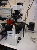 (1041464 bytes) == {{int:filedesc}} == {{Information| |Description = [[Inverted microscope]]. Nikon TE2000S. |Source={{own}} |Date = 2007-09-25 |Author = Nuno...(1,944 × 2,592 (1,017 KB)) - 14:53, 7 September 2020
 512×512 (27246 bytes) {{Information |Description = An image from an inverted microscope using a 40X objective illustrating raphides along with a lot of debry...(512 × 512 (27 KB)) - 14:40, 14 November 2020
 DescriptionNikon-Inverted fluorescence microscope.jpg English: A Nikon Inverted fluorescence microscope in laboratory. Date 5 December 2016, 11:19:07 Source...(3,264 × 2,448 (1.71 MB)) - 19:32, 14 June 2020
 DescriptionOlympus Gemini Inverted Microscope - NCMIR.jpg English: Olympus Gemini Inverted Microscope - NCMIR Date 3 March 2008 Source https://picasaweb...(1,063 × 1,600 (227 KB)) - 20:10, 20 October 2020
 DescriptionInverted Microscope, MNIT.jpg English: Inverted microscope from MNIT, Jaipur Date 9 January 2021 Source Own work Author Nikunj12387...(4,608 × 2,176 (2.23 MB)) - 17:02, 20 January 2021
 DescriptionBiologist using inverted microscope 01.jpg Português: Bióloga estudante de mestrado estudando células de câncer dentro de um frasco, com auxílio...(3,456 × 2,304 (2.6 MB)) - 13:07, 23 November 2023
 DescriptionFlask of cancer cells on inverted microscope.jpg Português: Frasco com células de câncer de pele sendo estudadas por uma bióloga com uso de...(2,601 × 1,729 (1.07 MB)) - 04:23, 28 October 2020
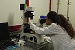 DescriptionBiologist using inverted microscope 02.jpg Português: Bióloga estudante de mestrado estudando células de câncer dentro de um frasco, com auxílio...(3,456 × 2,304 (2.83 MB)) - 13:07, 23 November 2023
 DescriptionBiologist using inverted microscope 06.jpg Português: Bióloga estudante de mestrado estudando células de câncer dentro de um frasco, com auxílio...(3,456 × 2,304 (2.15 MB)) - 13:07, 23 November 2023
 DescriptionBiologist using inverted microscope 05.jpg Português: Bióloga estudante de mestrado estudando células de câncer dentro de um frasco, com auxílio...(3,456 × 2,304 (2.53 MB)) - 13:07, 23 November 2023
 DescriptionBiologist using inverted microscope 07.jpg Português: Bióloga estudante de mestrado estudando células de câncer de pele de rato, linhagem B16F10...(3,456 × 2,304 (2.78 MB)) - 13:07, 23 November 2023
 DescriptionBiologist using inverted microscope 04.jpg Português: Bióloga estudante de mestrado estudando células de câncer dentro de um frasco, com auxílio...(3,456 × 2,304 (2.65 MB)) - 13:07, 23 November 2023
 DescriptionTrinocular-Inverted-Metallurgical-Microscope.jpg English: Trinocular Inverted Metallurgical Microscope. Date 13 June 2016 Source Sathish Rao...(600 × 291 (98 KB)) - 18:38, 21 February 2024
 DescriptionKöhler Illumination with the Inverted Microscope (15174751101).jpg Ask your ZEISS account manager for a lab poster! You'll find more knowledge...(3,000 × 4,200 (638 KB)) - 09:08, 15 July 2024
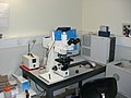 (2269351 bytes) By Richard Wheeler ([[User:Zephyris|Zephyris]]) 2007. [[Microscope]] with [[mercury bulb]] for [[fluorescence microscopy]], attached to a...(3,072 × 2,304 (2.16 MB)) - 05:31, 20 May 2023
 2007-05-09 23:17:46 | SubtleGuest | 1016368 | 2816×2112 | Image taken with Canon Powershot S3 on Nikon TS100 inverted microscope by the author. English...(2,816 × 2,112 (993 KB)) - 16:01, 18 October 2020
 4.0 truetrue English A slide of stained tissue, analyzed at inverted digital microscope Russian Окрашенный срез тканей исследуется на цифровом инвертированном...(4,288 × 2,848 (6.85 MB)) - 17:54, 3 December 2022
 Taken with Olympus cellStandard software for Olympus inverted microscope Location- University of Southern California, Los Angeles I, the copyright holder...(635 × 425 (90 KB)) - 05:14, 19 September 2020
 Other information English: Taken on a Zeiss inverted microscope with 40X phase contrast optics. I, the copyright holder of this work, hereby publish it...(1,648 × 1,692 (230 KB)) - 19:11, 10 October 2020


.jpg)


