
Sebaceous hyperplasia
This article needs additional citations for verification. (June 2009) |
| Sebaceous hyperplasia | |
|---|---|
 | |
| Sebaceous hyperplasia in 55-year-old woman, diagnosis was histologically verified. | |
| Specialty | Dermatology |
Sebaceous hyperplasia is a disorder of the sebaceous glands in which they become enlarged, producing flesh-colored or yellowish, shiny, often umbilicated bumps on the face.[1] Newly formed nodules often swell with sweating (which is pathognomonic for the condition), but this diminishes over time.
Sebaceous glands are glands located within the skin and are responsible for secreting an oily substance named sebum. They are commonly associated with hair follicles but they can be found in hairless regions of the skin as well. Their secretion lubricates the skin, protecting it from drying out or becoming irritated.[2]
Sebaceous hyperplasia generally affects newborns as well as middle-aged to elderly adults. The symptoms of this condition are 1–5 mm papules on the skin, mainly on the forehead, nose and cheeks, and seborrheic facial skin. The papules may be cauliflower-shaped. In infants, acne is sometimes associated with sebaceous hyperplasia.
Additional photos
-
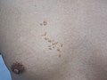 Photography of sebaceous hyperplasia, showing a group of papules, in this case on the chest with linear distribution pattern.
 -
 Dermoscopy of sebaceous hyperplasia. Note the aggregation of yellowish-white clods with linear vessels between or above the clods.
 -
 Dermoscopy of sebaceous hyperplasia with digital high dynamic range. Note the multi-lobulated clods with central openings.
 -
 H&E staining of biopsied lesion of sebaceous hyperplasia: Note the multiple, mature sebaceous lobules attached to the central dilated duct in the upper dermis.
 -
 Sebaceous hyperplasia, lateral right temple marked for biopsy with adjacent malignant melanoma in situ, evolving, medial right temple
 -
 Sebaceous hyperplasia with surrounding chronic folliculitis, right mid chest


See also
References
- ^ James, William D.; Berger, Timothy G.; et al. (2006). Andrews' Diseases of the Skin: Clinical Dermatology. Saunders Elsevier. p. 662. ISBN 978-0-7216-2921-6.
- ^ Jo Ann Coers Eurell; Brian L. Frappier (2006). Dellmann's textbook of veterinary histology. Wiley. p. 29. ISBN 9780781741484.