
File:Chlamydia pneumoniae.jpg
No higher resolution available.
Chlamydia_pneumoniae.jpg (275 × 180 pixels, file size: 7 KB, MIME type: image/jpeg)
| This is a file from the Wikimedia Commons. Information from its description page there is shown below. Commons is a freely licensed media file repository. You can help. |
Summary
| Description |
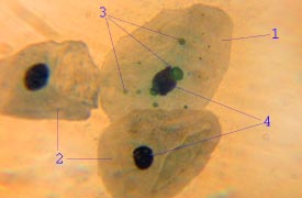
English: Chlamydia pneumoniae in epithelial cell. Acute bronchitis. 1 - infected epitheliocyte, 2 - uninfected epitheliocytes, 3 - chlamydial inclusion bodies in cell, 4 - cell nuclei. Micrograph
Русский: Chlamydia pneumoniae в клетке плоского эпителия при остром бронхите. 1 - инфицированная клетка, 2 - неинфицированные клетки, 3 - тельца включения C.pneumoniae в инфицированной клетке, 4 - ядра клеток. Микрофотография орашенного мазка. Объектив 100x (масляная иммерсия). Способ окраски - согласно пат. РФ № 2281472 |
||
| Date | |||
| Source | Own work | ||
| Author | Eutensist | ||
| Permission (Reusing this file) |
|
File history
Click on a date/time to view the file as it appeared at that time.
| Date/Time | Thumbnail | Dimensions | User | Comment | |
|---|---|---|---|---|---|
| current | 07:00, 15 January 2010 | | 275 × 180 (7 KB) | Eutensist | {{Information |Description={{en|1=Chlamydia pneumoniae in epithelial cell. Acute bronchitis. 1 - infected epitheliocyte, 2 - uninfected epitheliocytes, 3 - chlamydial inclusion bodies in cell, 4 - cell nuclei. }} {{ru|1=Chlamydia pneumoniae в клетк |
File usage
The following pages on the English Wikipedia use this file (pages on other projects are not listed):
Global file usage
The following other wikis use this file:
- Usage on ar.wikipedia.org
- Usage on arz.wikipedia.org
- Usage on cs.wikipedia.org
- Usage on de.wikipedia.org
- Usage on en.wiktionary.org
- Usage on es.wikipedia.org
- Usage on fr.wikipedia.org
- Usage on gl.wikipedia.org
- Usage on it.wikipedia.org
- Usage on nl.wikipedia.org
- Usage on pl.wikipedia.org
- Usage on pt.wikipedia.org
- Usage on ru.wikipedia.org
- Usage on sr.wikipedia.org
- Usage on sv.wikipedia.org
- Usage on tr.wikipedia.org
- Usage on uk.wikipedia.org
- Usage on www.wikidata.org